IV. First results and remarks
Labeling the head tetrahedra with labels BRAIN, SKULL or SCALP
1. Results
We can see in the following figures the results (exteriors surfaces)
of our labeling algorithm, first
for the scalp, then for the skull and finally for the brain.
In figures concerning the brain, we can see that the set B is
smaller than the set in the MRI volume. The reason is the size of the
tetrahedra, since the length of an edge of a tetrahedron belonging
to the ART volume for n=4 is close to the thickness of
the skull.
This problem will disappear with a resolution level of the
ART resolution n higher than 4 (see
Results with n=5).
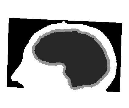
Initial segmented MRI volume.
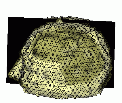
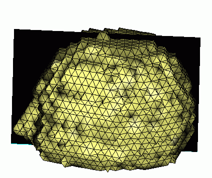
External surface of the scalp.
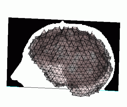
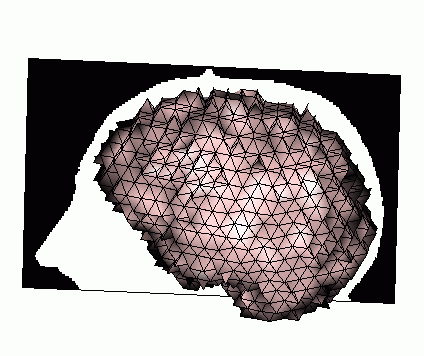
External surface of the skull.
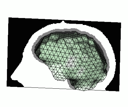
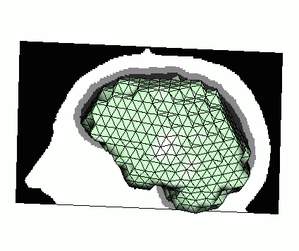
Surface of the brain.
2. Remarks
a) The method presented here is adaptable for more than 3 components
as long as they have the same structure, i.e. imbricated spheres.
For example we can imagine to use the same strategy to label the head
with SCALP, SKULL, CEREBRO-SPINAL FLUID and BRAIN as labels.
b) The choice of the thresholds mu is very
important for the quality
of the results. In fact, the conservation of the different
topologies is guaranteed by the use of simple tetrahedra, but
the thicknesses and the accurates of the tetrahedral tissues components
strongly depend on these thresholds.
I. Introduction
II. Topological tools
III. Method to label the head
IV. First results and remarks
V. Results with n=5
VI. Numerical values
VII. Visible Human with CSF
Main page


