Homotopic labeling of head tetrahedra
I. Introduction
1. Goal
In applications based on Electroencephalography (EEG) and
Magnetoencephalography (MEG), electromagnetic field propagation is
numerically calculated, for example using finite element methods.
This computation is based on a meshed model of the head tissues.
In this site, we propose a method to obtain such head models.
In our model, the head will be composed of identical
tetrahedra, each tetraheron being labeled with the corresponding anatomical
tissues: BRAIN, SKULL...
Our labeling is done under topological constraints in order to
respect the topological arrangement of the head, i.e. interwoven spheres.
We first present a method to decompose a part of the space into
tetrahedra (see next paragraphs). Then, we introduce the notion of
simple tetrahedron that allows us to propose the definition
of homotopic deformations (see Topological
tools).
Finally, we explain the method we use to label the tetrahedra of the
head using the homotopic deformations (see
Method to label the head); some results and comments are also presented
(see Results and remarks,
Results with n=5,
Numerical values).
2. Material
The data we have consist of a MRI volume representing a human head, and this
image is segmented to extract anatomical tissues. Then this
segmentation will be used for the labeling of the tetrahedra of the
space at the following steps.
We do not explain here in details the process used to obtain the segmented
MRI from the initial image as it is not the goal of this site.
A report of Elena Martinez-Perez (.pdf file) containing more explanations
about the segmentation step can be downloaded here:
report.
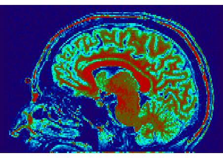
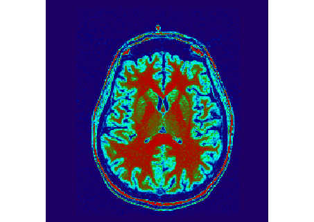
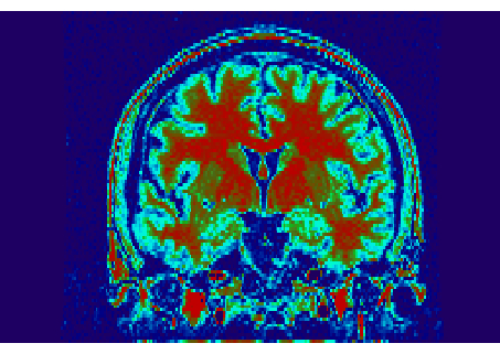
Initial MRI volume.
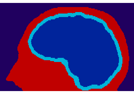
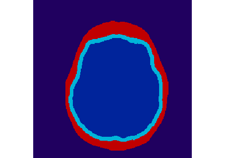
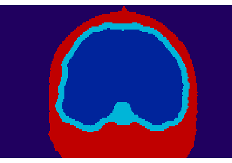
Segmented MRI volume:
dark blue <-> background, red <-> scalp,
sky blue <-> skull, blue -> brain.
3. Tetrahedral decomposition of the space
We use a specific model called Almost Regular Tessalation (ART)
volume mesh, based
on the work of J. Pescatore during his PhD thesis.
In this model, a portion of the space (that is a polyhedron)
is composed of tetrahedra,
each tetrahedron being close to an
equilateral tetrahedron. This set of tetrahedra is obtained by
successive subdivisions of the space that is initially a polyhedron
P composed of 24
tetrahedra sharing a same vertex. This initial polyhedron contains
the segmented MRI volume obtained before.
At each step, each tetrahedron t composing P
is subdivided into 8 tetrahedra congruent to t
scaled by a factor 1/2.
This process is proved to be subdivision invariant.
The number and the size of the obtained tetrahedra
depend on the number n of successive subdivisions.
The ART volumes
presented here are obtained after n=4 subdivisions,
and so the space
is composed of 24*(8^n) = 98304 tetrahedra.
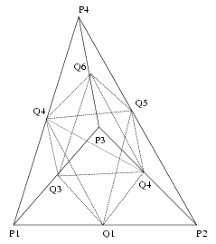
Subdivision of a tetrahedron
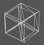
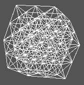
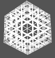
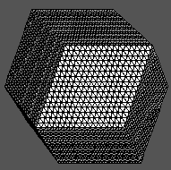
Successive subdivisions of the 24 initial tetrahedra representing
the space and containing the head,
with from left to right: n=0
(24 tetraedra), n=1 (192 tetraedra), n=2 (1536 tetraedra)
and n=4 (98304 tetraedra).
Based on this tetrahedral representation of the space,
first we have to label the tetrahedra composing the head, and after
we have to label each tetrahedron of the head with the corresponding
anatomical tissue, from the MRI segmented volume.
I. Introduction
II. Topological tools
III. Method to label the head
IV. FIrst results and remarks
V. Results with n=5
VI. Numerical values
VII. Visible Human with CSF
Main page


