Labeled tetrahedral mesh of a child MRI - n=5
Here we can see images obtained from a child MRI, with n=5
(this segmentation is obtained by Dr Najib Gadi, thanks to a grant from the ministry or research: RNRT Adonis).
A particularity of the child MRI is that the structures may be
very thin (see the last images). Our method allows us to recover
a minimal layer of
each structure in the labeled mesh.
In the example, the little labeled components have been removed.
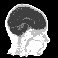

Left: MRI and right: labeled tetrahedral mesh.

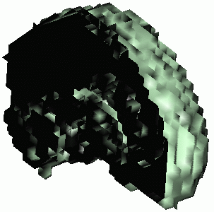


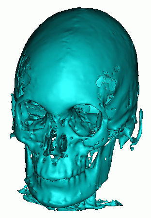
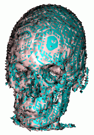
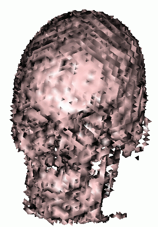
The surface of the segmented skull (left), superposed with the tetrahedral
mesh (middle) and the tetrahedral mesh alone (right).
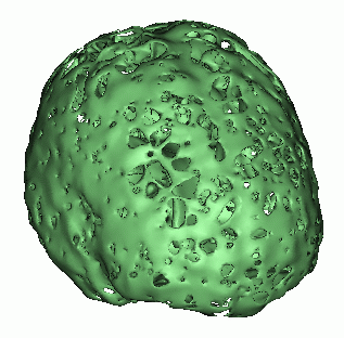

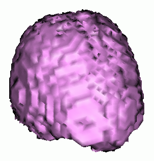
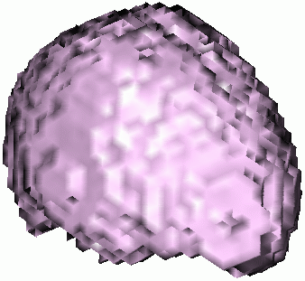
The surface of the segmented CSF (left), superposed with the tetrahedral
mesh (middle) and the tetrahedral mesh alone (right).
I. Introduction
II. Topological tools
III. Method to label the head
IV. FIrst results and remarks
V. Results with n=5
VI. Numerical values
VII. Visible Human with CSF
Main page