|
|
|
|
|
|
|
|
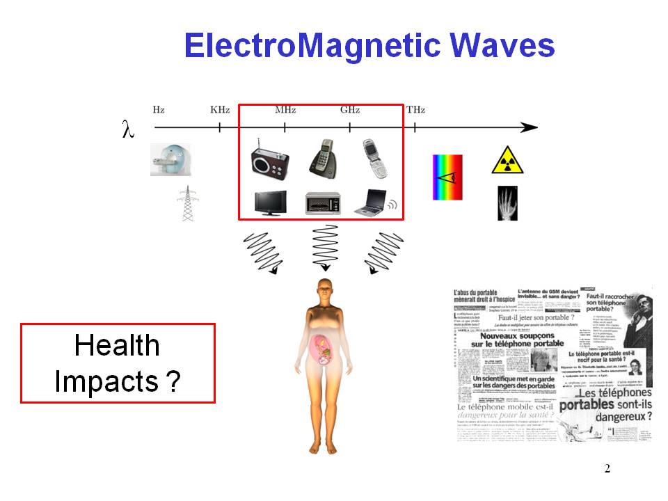
Several international scientific organizations and institutes, such as the World Health Organization (WHO) and the COST 281, have expressed the need for developping numerical models of the human body to enable precise studies of the interactions between radio-frequency electromagnetic waves and biological tissues. Several previous works have focused on developing models of adults and children heads, based on magnetic resonance imaging (MRI) data. New usage habits of mobile phones (hand free kits, ...) and the introduction of new technologies based on electromagnetic fields (Wifi, ...) have raised the need to develop whole body numerical models, based on MRI and computerized tomography (CT) data. With the advent of obstetrical imaging, models of the fetus and the pregnant woman are now considered.
 |
This project, entitled FEMONUM (FEtus and MOther NUmerical Models), aims at providing numerical models generated from a large database of imaging exams. The proposed models, evaluated by an obstericians and a group of pediatric radiologists, will enable precise dosimetry studies on the complex, highly variable and evolving anatomy of the pregnant woman. |
This project was funded by the Fondation Santé et Radiofréquence. The numerical models will be made available to the scientific community, filling an important gap in the list of existing human body models.
The modeling approach is based on medical image segmentation and smooth surface mesh generation. Two types of medical imaging modalities are used to screen and model the fetus:
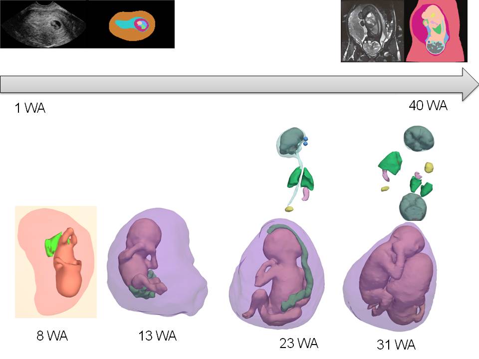
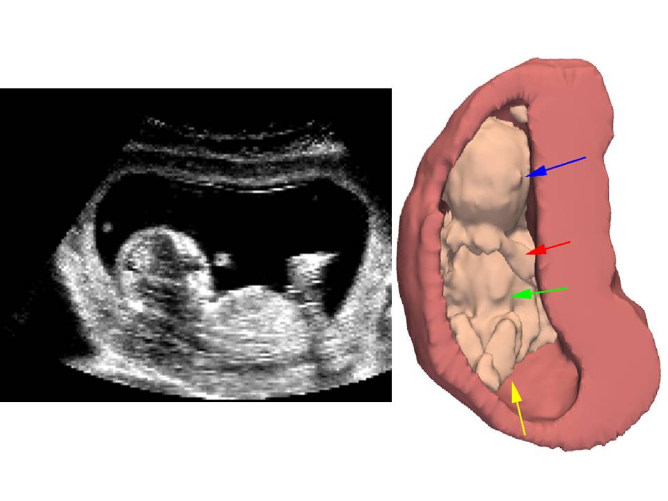
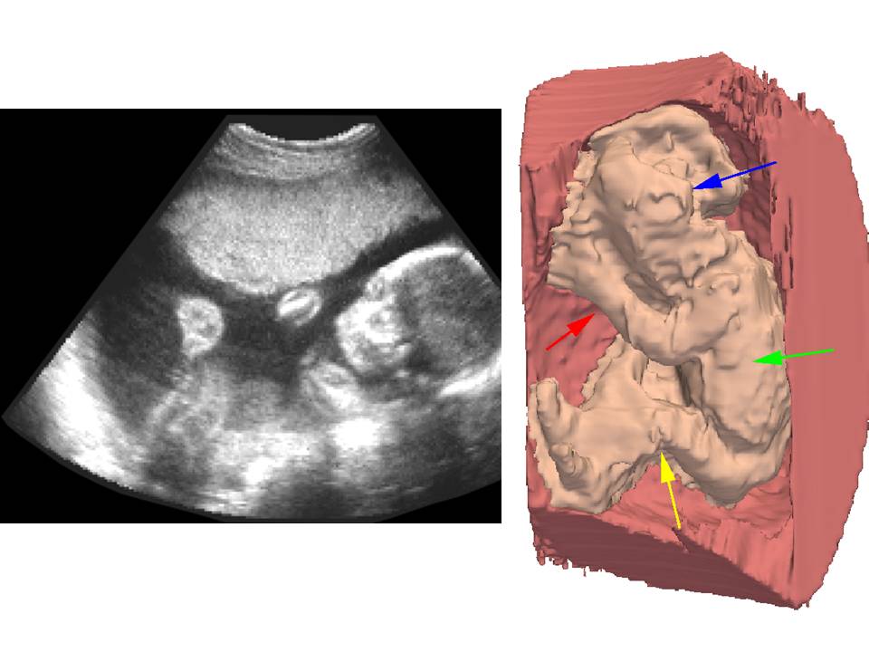
A database of 19 volumes of 3D ultrasound acquired between 8WA et 22 WA was provided
by collaborating obstetricians (Hospital Beaujon, Paris, FRANCE) and by Philips Healthcare.
Segmentation of the fetus body is performed in two steps:
 |  |
|
|
|
(1) field of view, (2) uterine internal wall. |
|
(3) uterine wall and fetus body, (4) fetus body. |
MRI image data provide a clear visualization of several maternal abdominal tissues (skin, bone, fat, muscles,...) et of fetal organs (body envelope, brain, lungs, heart,...).
In collaboration with the pediatric radiology department of the hospital Saint Vincent de Paul (Paris, France), we first performed a study to define the best suited MRI protocol for the segmentation and modeling of the fetus. The Steady State Free Precession (SSFP) sequence was selected, offering the best image quality for the segmentation task. Results of this study were published in a paper.
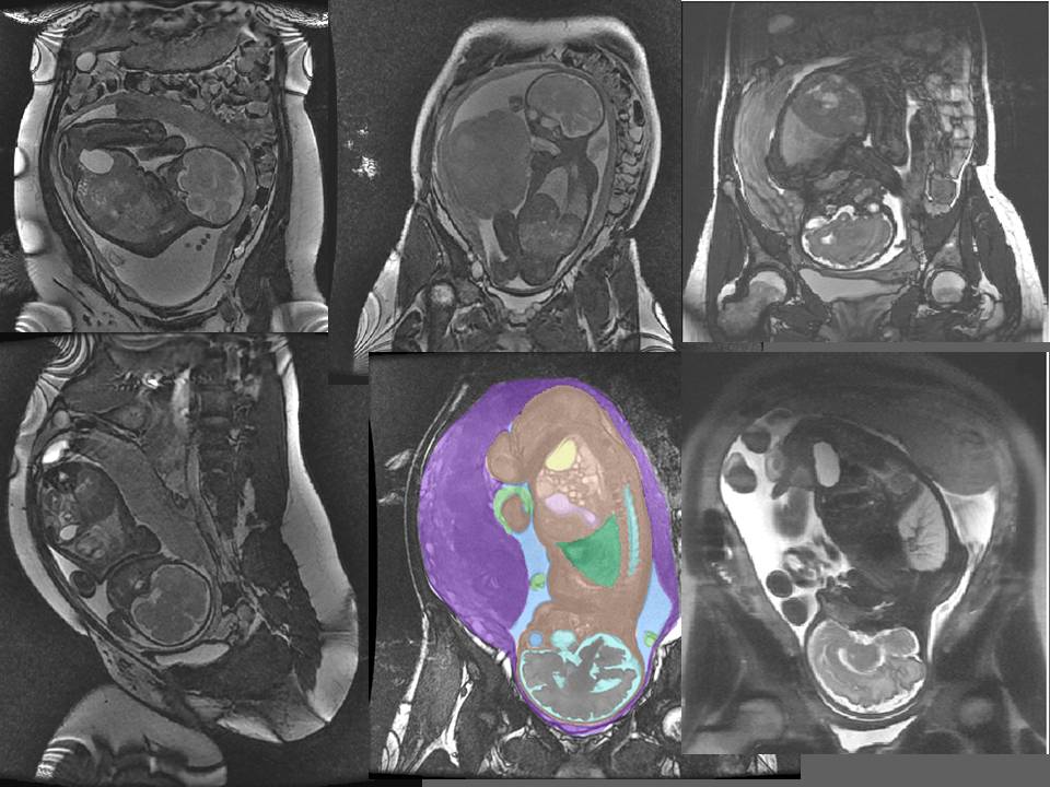 |
| Examples of SSFP & FFSE MRI data of fetuses. |
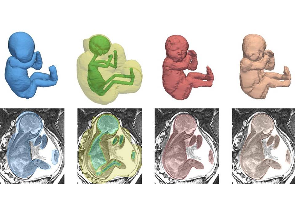
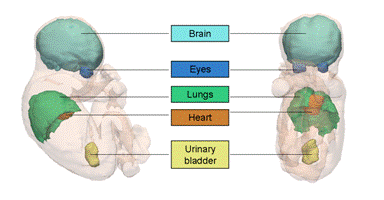
A methodological framework was developed for automated segmentation of several fetal structures, including the eyes, the brain-skull content, the spinal cord, the urinary bladder, and the lungs. The segmentation process is based on the extraction of landmark points based on appearance models, iterative orientation of the fetus based on the landmark points and fine segmentation of tissue interfaces with a contrast-sensitive graph-cut partitioning of image data, within narrow bands of image data.
.
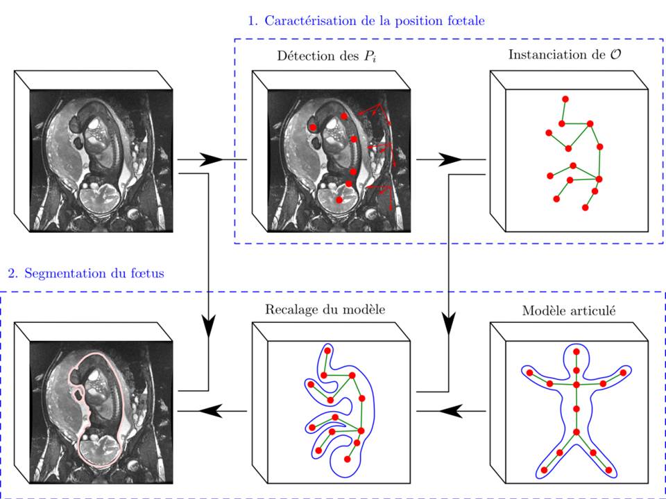 | 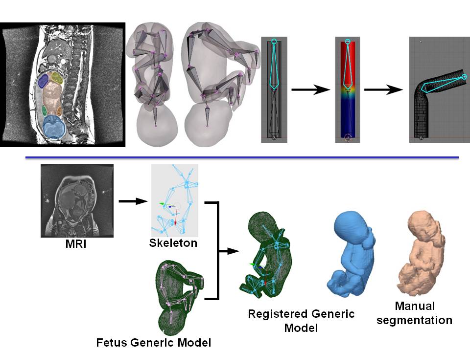 |
| Extraction of Landmark Points on the Foetal Skeleton | Registration of a Generic Foetal Articulated Model on the Skeleton |
|
|
|
|
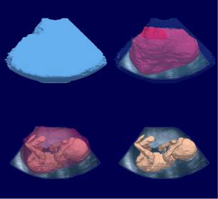
Final Segmentation of the Foetal Body Envelope with Graph-Cuts |
Refinment of the 3D Fetus Segmentation with Internal Organs |
 |
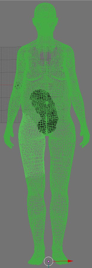
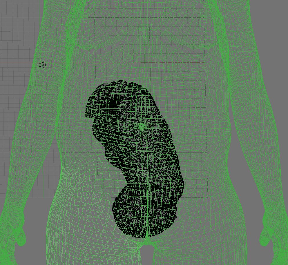
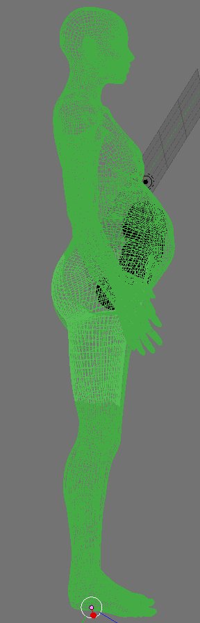
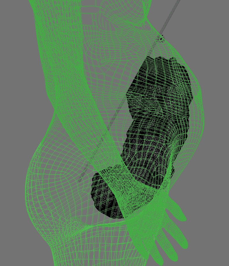
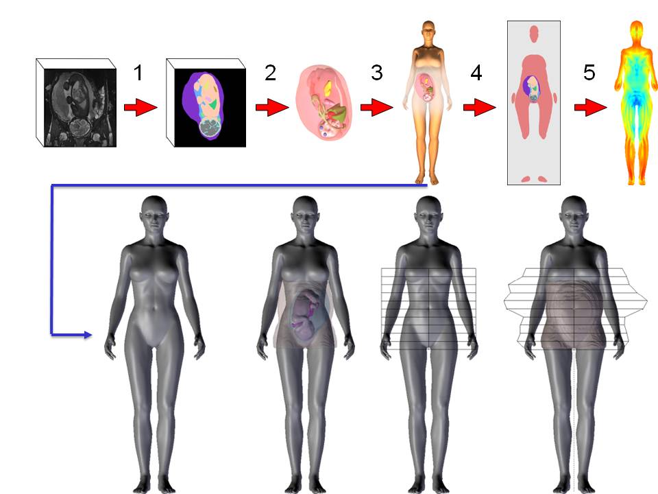
Victoria is a non-gravid woman body envelope developed by Daz Studio, which can be manipulated and animated with the software tool Blender.
We used Victoria's envelope to generate hybrid models of pregrant women:
 |
|
|
|
|
|
|
Mesh-based pregnant woman model (fetal model based on MRI data) |
|||
A pixel-based model is finaly generated, at a desired spatial resolution, with different labels assigned to the different tissues.
|
|
|
|
|
|
Pixel-based pregnant woman model |
|||