

|
|
Medical Imaging | |
| Département TSI | CNRS UMR 5141 LTCI |
Cardio-vascular imaging:
Brain imaging:
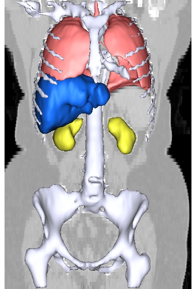
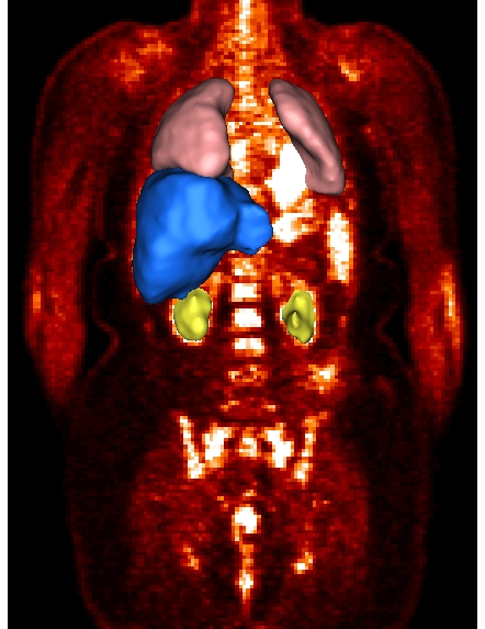
Thorax imaging:
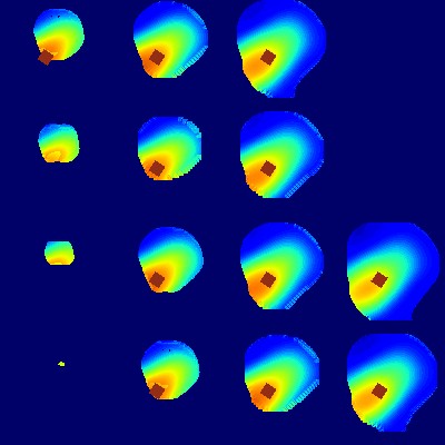
Regularized reconstruction of 3D MRA images
of high resolution from images of anisotropic resolution
PhD thesis of Elodie Roullot (in collaboration with Alain Herment and
Elie Mousseaux, INSERM)
 |
 |
|||||
| Two corresponding slides fron the anisotropic acquisitions. | ||||||
 |
 |
 |
||||
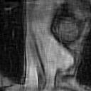
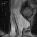
| Reconstruction without constraints | Reconstruction with smoothness constraints, but without contour preservation | Reconstruction with smoothness and contour preservation constraints | ||||
 |
 |
 |
| 3D MRI acquisition | Modeling using superquadrics | Volume of the left ventricle during the cardiac cycle |
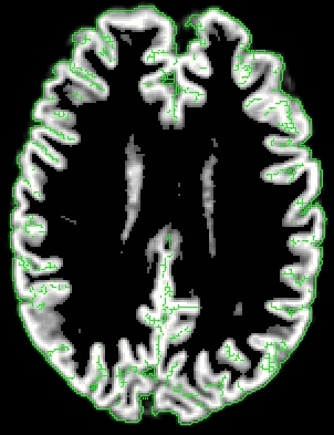
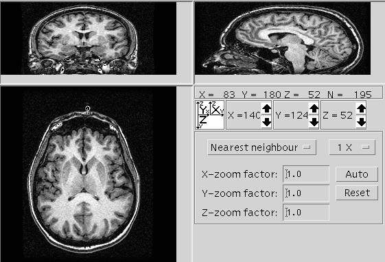
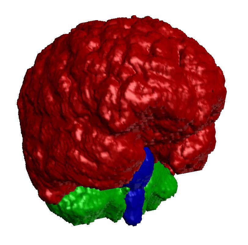 Segmentation of the brain,
the brainstem and the cerebellum
Segmentation of the brain,
the brainstem and the cerebellum
 Segmentation
of the skin, the muscles, the skull, the brain, the cerebellum, the brainstem
Segmentation
of the skin, the muscles, the skull, the brain, the cerebellum, the brainstem
 |
 |
 |
 |
 |
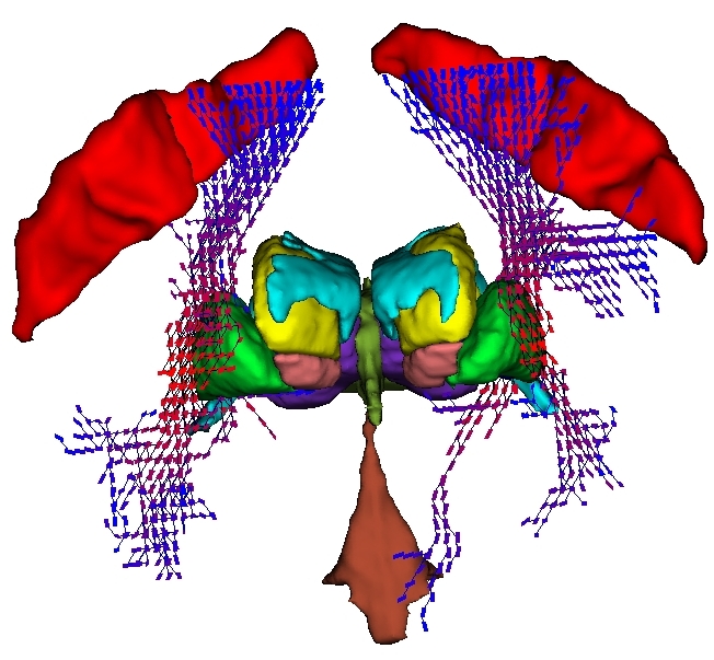
| Contrainsts with respect to already recognized structures | Approximate location provided by the atlas | Information provided by the grey levels of the image | Relative position with respect to recognized structures | Relative position with respect to recognized structures (3D view) |
 |
 |
 |
 |
| Superimposition of the atlas and the image before recognition of the caudate nucleus | Approximate location provided by the atlas | Fusion of informations | Segmentation and recognition of the caudate nucleus |
 |
 |
| Of few structures of the atlas | Structures recognized in the 3D image |
 |
 |
 |
|
Multi-scale analysis of functional images (TP and fMRI) and graph matching |
Superimposition of activation areas during a primary motor task (right hand) on the subject anatomy | Localization of an activation due to a vibration in the hand with respect to two cortical sulci |
 |
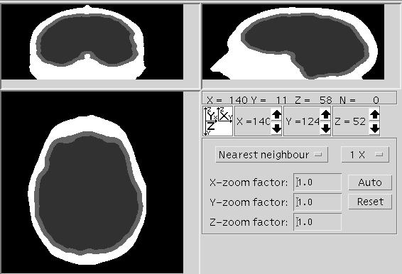 |
| 3D MRI Image | Segmentation of the skin, the skull, the brain |
 |
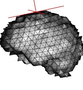 |
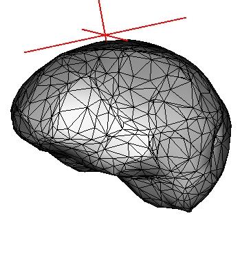 |
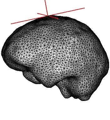
| High resolution mesh | Surface of a tetraedral mesh | Smoothed mesh |
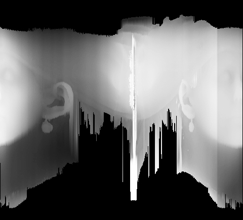
Acquisition of a head range image, triangular mesh after filtering
constituting a head model.
Morphological measurements on the ear.
Average ear included in a generic phantom.
Voxel representation used for numerical simulations of field propagation,
superimposition of voxel components on the mesh.
Maximum SAR (``specific absorption rate'') in a cube of 10 gramms.
 |
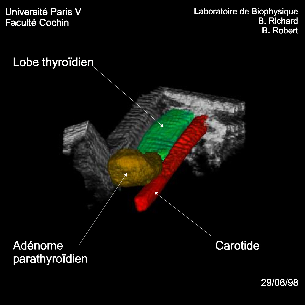 |
| 3D reconstruction of a foetus | 3D reconstruction of carotid and thyroid |
Non linear matching in oncology
PhD theses of Oscar Camara and Gaspar Delso, with a contribution by
Olivier Colliot (in collaboration with Jean-François Stévenet
and Sylvie Hammer, Segami, Hervé Foehrenbach, Val-de-Grâce
Hospital, Xavier Marchandise and Jean-Yves Rousseau, CHRU Lille, Jean-Pierre
Rigo, Monaco hospital)