
Spatio-temporal analysis of pediatric magnetic resonance images


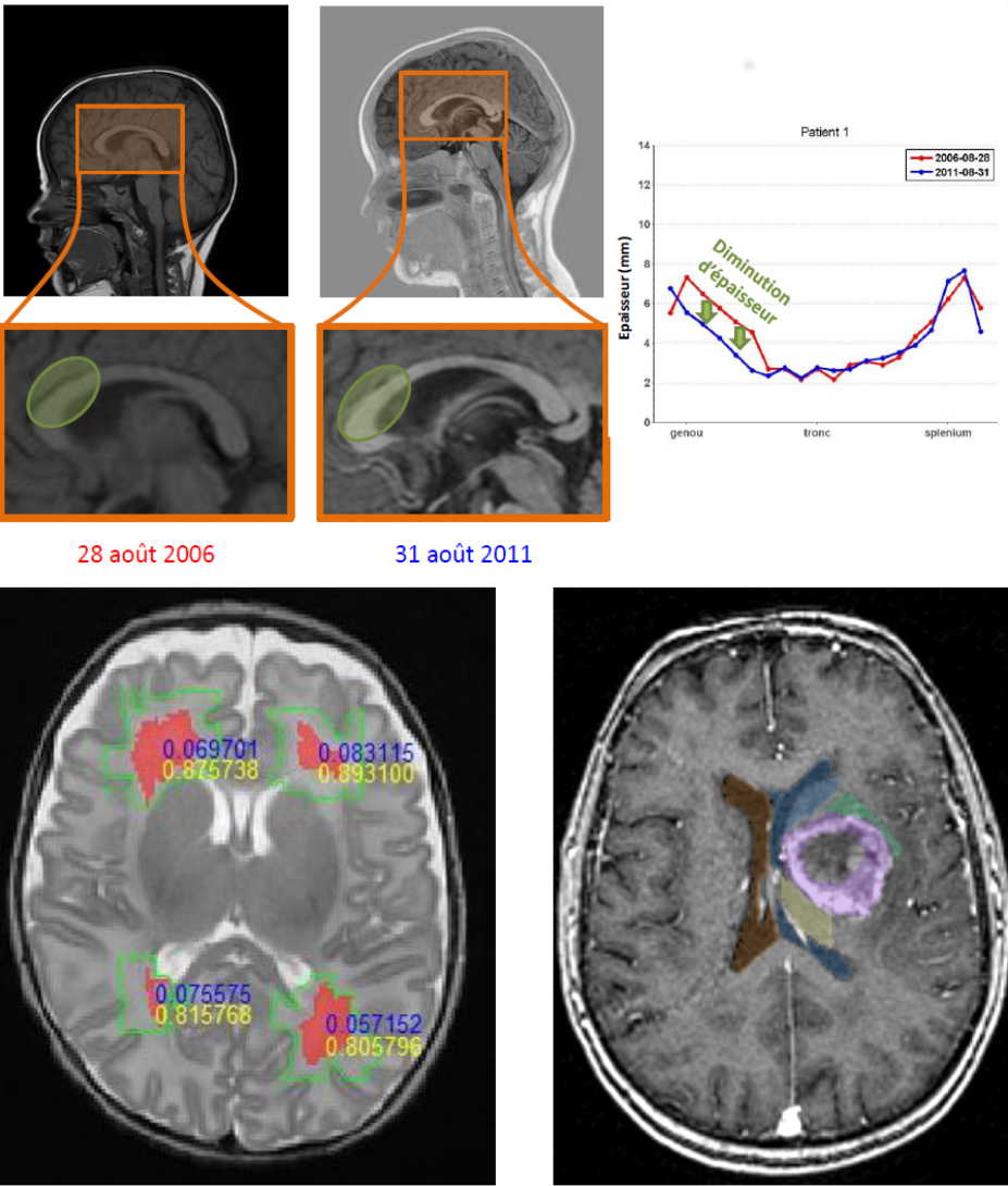
Project description: The advances in medical imaging require to develop quantitative or semi-quantitative methods to improve accuracy in the image analysis results. Advances in medical image analysis provide such tools, but there is still an important gap regarding pediatric brain imaging, even though there is an increasing medical demand. This project aims at contributing to fill this gap, focusing on brain magnetic resonance imaging (MRI) of infants, newborns and premature babies, which raise specific issues due to the particular grey/white matter contrast related to the physiological myelination process, the very fast but not continuously observed evolution of the brain structures and possible pathologies, and the high intra-and inter-subjects variability. One of these issues is that the data at hand are noisy, ambiguous, scarce in nature and sparse in time. In turn, expert medical knowledge is available, but is prone to change and evolution. From this point of view the project tackles one of the very cutting edge questions in data analysis, that is how to extract and understand meaningful patterns where the data are scarce but expert knowledge, continuously enriched, is available. We propose to develop structural representations of knowledge and image information in the form of graphs and hypergraphs, which will be exploited to guide spatio-temporal image understanding (segmentation, recognition, quantification, comparison over time, description of image content and evolution). The aim is to aid diagnosis, pathology analysis and patients' follow-up. Applications will include the analysis of hyperintensities on the white matter, the volumetry of corpus callosum and its evolution, and neuro-oncology with the study of the influence of tumors on surrounding structures over time. The project involves specialists in medical image analysis, structural knowledge representation and pediatric neuro-imaging.